Lumbosacral Disease
Why Should I Bring my Pet to Willows for Treatment of Lumbosacral Disease?
Willows is one of Europe’s leading small animal referral centres. Our state-of-the-art hospital is led by internationally renowned Specialists, committed to providing the highest standards of veterinary care. Our team of Specialist Neurosurgeons have extensive experience, having treated hundreds of dogs with lumbosacral disease, providing gold standard management and treatment for all our patients.
Our Neurology and Orthopaedic Specialists are supported by our multi-disciplinary team of Specialists across a number of different disciplines including; Anaesthesia, Diagnostic Imaging and Emergency and Critical Care. Willows also has a large dedicated team of Vets, Nurses, Physiotherapists and clinical support staff available 24 hours a day, every day of the year to provide the best possible care for your pet.
What is the Lumbosacral Disc?
The lumbosacral disc is the last disc in the lower back between the last bone in the spine (lumbar vertebra) and the bone that supports the pelvis (sacrum).
What are the Signs of Lumbosacral Disease?
Back pain is the key clinical feature of the disease. Typically, affected animals are reluctant to jump and climb, and they may have difficulty rising from a lying position. Groaning or spontaneous yelping may be a feature. Muscle wastage of the hamstrings is also common. Less common signs include back leg lameness (due to spinal nerve compression), incontinence and reduced tail movement.
How does Thoracolumbar Disc Disease Occur?
Lumbosacral stenosis is a neurological condition in which nerves at the base of the spine are compressed by a bulging disc or other tissues. Ageing may result in dehydration and degeneration of this disc which may then bulge (protrude) and compress or ‘trap’ regional nerves. The resultant narrowing of the canal in the spine (the vertebral canal) or the exit holes between the bones (the vertebral foramen) is referred to as stenosis. The underlying cause of the disc degeneration and bulging is unclear, although in some dogs it may be associated with instability of the bones of the spine.
Large breeds of dogs are more commonly affected with lumbosacral stenosis, and German Shepherd Dogs and Border Collies appear to be particularly prone to the condition. It is an uncommon complaint in cats.
How is Lumbosacral Disease Diagnosed?
The history and examination are an extremely important part of the diagnosis of lumbosacral disease. MRI scanning is the best method for confirming the presence of many spinal conditions, including lumbosacral stenosis. MRI scanning uses high powered magnets and a computer to generate images of the spine (this is the same technique and the same equipment which is used for body scanning in humans). It provides detailed information on the location and extent of any soft tissue compressions of the spinal nerves in the lumbosacral spine.
How can lumbosacral disease be treated?
The two principle methods of managing thoracolumbar disc disease are:
Conservative treatment: As in people with back pain associated with a ‘slipped disc’ in the lower back, the majority of dogs and cats with lumbosacral stenosis can be successfully managed without the need for surgery. It is often necessary to modify exercise to avoid strenuous activities that involve jumping, climbing, twisting and turning. Dogs should initially be walked on a lead (short distances, frequently) and exercise should be gradually increased over a number of weeks. Overweight patients should be placed on a calorie restricted diet. The majority of affected animals will benefit from receiving pain killing medications. Anti-inflammatory agents, neuropathic drugs and muscle relaxants may all be beneficial. Physiotherapy and hydrotherapy can also be useful treatments. Lumbosacral stenosis may also be managed by injecting a long-acting steroid (cortisone) around the compressed spinal nerves via a lumbar puncture. Repeat injections may be necessary in some patients. The technique necessitates a general anaesthetic.
Surgery: Some patients with lumbosacral stenosis require surgery in order to relieve pain, back leg lameness and other problems such as incontinence. Various techniques may be performed, and these aim to remove pressure on compressed nerves and/or stabilise the lumbosacral spine. Removing bone from the top of the spine is referred to as a laminectomy, whilst enlarging the exit holes between the bones (the vertebral foramen) is referred to as a foraminotomy. Bulging disc material and other soft tissues that are compressing spinal nerves are removed. The bones surrounding the lumbosacral disc may be stabilised by the insertion of screws or pins that are secured with metal bars or cement. At Willows we have pioneered the use of custom-made 3D printed guides for this purpose.
What Can I Expect if my Dog is Treated for Lumbosacral Disease?
In conservatively treated pets, once signs have improved or have been resolved, it is often possible to gradually increase exercise and reduce or stop medications. ‘Lifestyle’ changes may be necessary e.g. it may be necessary for the activity of an agility dog to be modified. As in people, flare-ups are not uncommon, and these may necessitate further periods of medication and restricted exercise.
In surgically treated pets, a few days of hospitalisation is typically needed for pain relief. These animals are often subsequently treated in exactly the same way as conservatively treated pets. Most respond favourably following appropriate postoperative rehabilitation, although recurrence of back pain can be a feature in some cases.
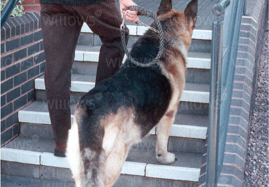
German Shepherd with lumbosacral stenosis that is reluctant to go up a small flight of steps due to back pain. Note also the dropped tail associated with the spinal nerve compression.
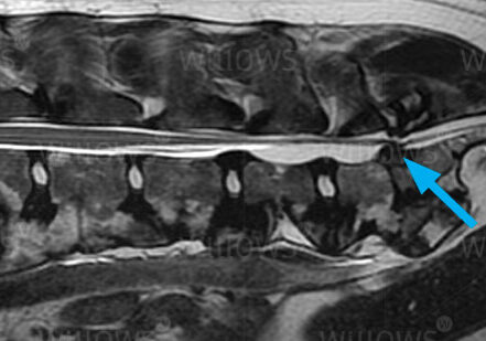
MRI scan of the spine showing lumbosacral stenosis due to a bulging disc (arrow)
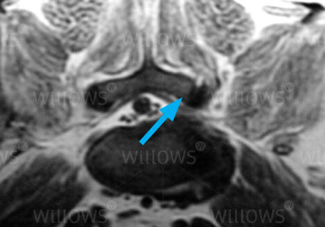
MRI scan of spine (cross section) showing lumbosacral stenosis due to a protruding (bulging) disc that is compressing the nerves on this side of the spine (arrow)
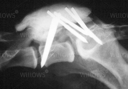
X-ray following lumbosacral stenosis surgery showing multiple pins embedded in cement that were used to stabilise the spine
To save this page as a PDF, click the button and make sure “Save as PDF” is selected.
Neurology
Find out more
To assist owners in understanding more about Neurological conditions, investigations and treatment we have put together a range of information sheets to talk you through the some of the more common neurological conditions seen by our Specialists.