Patellar Luxation
Why Should I Bring my Pet to Willows for Treatment of Patellar Luxation?
Willows is one of Europe’s leading small animal Orthopaedic referral centres treating over 1000 new patients a year. Our state-of-the-art hospital is led by internationally renowned Specialists committed to providing the highest standards of care. Our team of Orthopaedic Specialists have considerable collective experience of treating both dogs and cats who have patellar luxation.
Our Orthopaedic Surgeons are supported by our multi-disciplinary team of Specialists across a number of disciplines including; Anaesthesia, Diagnostic Imaging and Emergency and Critical Care. Willows has a large dedicated team of Nurses and clinical support staff available 24 hours a day, every day of the year to provide the best possible care for your pet.
What is Patellar Luxation?
The patella (kneecap) is a small bone at the front of the knee (stifle) joint, positioned between the quadriceps muscle and a tendon that attaches it to the shinbone (tibia). This is termed the quadriceps mechanism. As the knee bends and extends the kneecap glides in a groove (trochlea) at the end of the thighbone (femur). Occasionally, the kneecap slips out of the groove; this is called luxation (dislocation) of the patella. The dislocation usually occurs towards the inside (medial aspect) of the knee, however, it can also dislocate towards the outside (lateral aspect) of the joint. Patellar luxation can affect dogs and cats.
What are the Most Common Causes of Patella Luxate?
The patella luxates because it and the quadriceps mechanism, are not properly aligned with the groove. The cause of the abnormal alignment is often quite complex, involving varying degrees of deformity of the thighbone and shinbone. Kneecap dislocation is common in certain breeds of dogs, such as Poodles, Yorkshire Terriers, Staffordshire Bull Terriers and Labrador Retrievers often with both knees affected, which suggest that the condition may be genetic. Dislocation of the kneecap due to trauma is uncommon. Occasionally kneecap dislocation can be associated with rupture of the cranial cruciate ligament.
What are the Signs of Patellar Luxation?
The signs of patellar luxation can be quite variable. A ‘skipping’ action with the hind leg being carried for a few steps is common. This occurs when the kneecap dislocates out of the groove and resolves when it goes back in again. If both kneecaps dislocate at the same time, dogs and cats can have difficulty walking, often with a crouched back leg action.
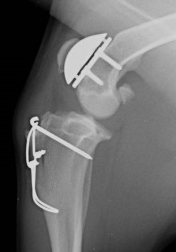
Fig 1: X-ray of a dog’s knee joint following a patella groove replacement
How is Patellar Luxation Diagnosed?
Examination may reveal muscle wastage (atrophy) in the affected leg(s). Manipulation of the knee may enable the detection of instability of the kneecap as it dislocates in and out of the groove. In some pets the knee-cap is permanently out of the groove. The severity of the dislocation is graded from one to four, four being the most severe. Whilst X-rays provide additional information, on the presence and severity of any associated knee osteoarthritis, it can be difficult to get X-rays that allow accurate assessment of the shape of the thighbone and shinbone. When possible a CT scan of the back legs is preferred to gain information regarding bone shape, which helps greatly in both surgical decision making and planning.
What Treatments are Available for Patellar Luxation?
Some pets with patellar luxation can be managed satisfactorily without the need for surgery. The smaller the pet and the lower the grade of luxation, the more likely it is that this approach will be successful. Exercise may need to be restricted. Hydrotherapy is often beneficial. Pets that are overweight benefit from being placed on a diet.
Many pets with kneecap dislocation benefit from surgery. The key types of surgery which may be required, include:
- Quadriceps realignment surgery: The aim of this surgery is to move a small piece of bone (the tibial tuberosity) at the top of the shinbone that is attached to the kneecap, and reposition it so that the kneecap is correctly aligned with the groove in the thighbone. The piece of bone is most often reattached with one or two small pins and wire. This procedure is called a tibial tuberosity transposition.
- Patella groove deepening surgery: If required this can be done by removing a block or wedge of bone and cartilage from the groove, deepening the base, and replacing the block or wedge. This makes the groove deeper, while at the same time preserving its smooth cartilage surface.
- Patella groove replacement: In some patients, particularly those who have significant osteoarthritis affecting the groove, or who have had previous unsuccessful kneecap dislocation surgery, can benefit from placement of a patella groove prosthesis, which resurfaces this part of the knee joint.
- Femoral osteotomy surgery: This involves changing the shape of the deformed thighbone by cutting it just above the knee and stabilising it in a new position with a bone plate and screws. This may be all that is needed to prevent the kneecap dislocating, however, additional procedures such as a tibial tuberosity transposition may also be required.
What can I Expect if my Pet is Treated for Patellar Luxation?
Exercise following surgery must be heavily restricted for the first few weeks during the healing process. After a few weeks, controlled lead exercise (on a lead) may be gradually increased and hydrotherapy may be recommended. X-rays are generally necessary between six and eight weeks following surgery to ensure bone healing is progressing without complication. Exercise may then be gradually increased in a controlled manner.
The outlook with kneecap dislocation surgery is generally good. Although all dogs and cats develop osteoarthritis of the knee to some degree, this is often not a cause of pain or lameness. Stiffness, especially after rest, can be a feature in some pets. Potential surgical complications include recurrence of the knee-cap dislocation and loosening of implants. These are uncommon.
To save this page as a PDF, click the button and make sure “Save as PDF” is selected.
Orthopaedics
Find out more
To assist owners in understanding more about Orthopaedics we have put together a range of information sheets to talk you through the some of the more common orthopaedic conditions seen and treated by our Specialists.