Third Eyelid Gland Prolapse
Why Should I Bring my Pet to Willows for Treatment of Third Eyelid Gland Prolapse?
Willows is one of Europe’s leading small animal referral centres. Our state-of-the-art hospital is led by internationally renowned Specialists, committed to providing the highest standards of veterinary care. As leaders in the field, our eye Specialists have experience of microsurgical techniques as well as access to a superb range of microsurgical instrumentation.
Our Ophthalmologists are supported by our multi-disciplinary team of Specialists across a number of disciplines including; Anaesthesia, Diagnostic Imaging and Emergency and Critical Care. Willows also has a large dedicated team of Nurses and clinical support staff available 24 hours a day, every day of the year to provide the best possible care for your pet.
What is the Third Eyelid and Third Eyelid Gland?
Dogs have three eyelids, the third eyelid being an extra eyelid that sweeps back and forth across the surface of the eye providing protection and spreading the tear film. The third eyelid is also called the nictitating membrane. The tear film is produced by two glands, namely the third eyelid gland (nictitans gland) and the lacrimal gland. The third eyelid gland is attached to the base of the third eyelid where it is not normally visible.
What are the Most Common Causes of ‘Third Eyelid Gland Prolapse’?
The gland is held in its normal position at the base of the third eyelid by a small ligament which attaches it directly onto the bone of the eye socket. If the ligament breaks the gland becomes mobile and slips down (known as a prolapse) from its normal position becoming visible above the edge of the third eyelid.
A third eyelid gland prolapse usually affects both eyes but not necessarily at the same time. The second eye often becomes affected after an interval of a few weeks to months.
The third eyelid gland produces approximately 40% of the tear film and so has an essential role in keeping the surface of the eye wet. When in the wrong position the gland does not produce tears effectively causing the surface of the eye to become dry. If the surface of the eye is dry, the eye becomes uncomfortable and susceptible to developing conjunctivitis, abnormal discharges and corneal ulcers.
What are the Signs of Cherry Eye?
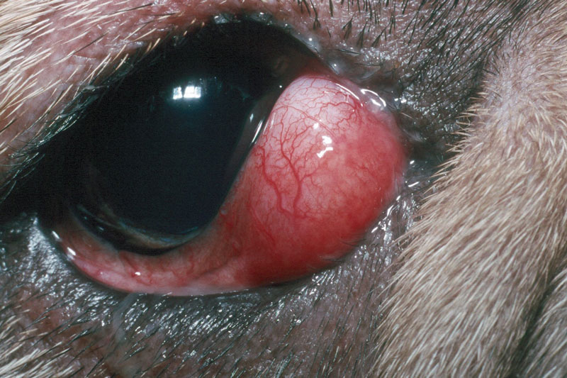
The prolapsed third eyelid gland is visible as a pink mass or ‘lump’ near the inner corner of the eye, resembling a cherry, hence the common term ’cherry eye’.
Third eyelid gland prolapse is much more common in dogs than in cats. It usually affects young dogs between the ages of six to twelve months, there is however some variation in the age of onset. Predisposed breeds include the Bulldog, Shih Tzu, Lhasa Apso, Cocker Spaniel, Great Dane and Mastiff.
Fig 1: A six month old English Bulldog with “cherry eye”
How is Cherry Eye Diagnosed?
This condition is typically diagnosed with a full ophthalmic examination assessing the typical clinical signs, consisting of a pink mass or ‘lump’ near the inner corner of the eye resembling a cherry, as mentioned above.
What Treatments are Available for Cherry Eye?
Surgery is recommended to return the gland to its normal position at the base of the third eyelid where it cannot be seen and can function normally. There are several surgical techniques that can be performed. At Willows the most common technique used is the ‘pocket’ technique.
Some breeds such as the Bulldog are predisposed to both ‘cherry eye’ and ‘dry eye’ conditions and it is therefore particularly important in such breeds that the prolapsed gland is replaced and not removed as this can lead to ‘dry eye’ later in life. If the gland is left untreated it will not produce tears normally and may become enlarged and red, causing discomfort.
What can I Expect if my Pet is Treated for Cherry Eye?
Your pet will stay in the hospital for the day and undergo a general anaesthetic in order for the ‘pocket technique’ to be performed. This technique involves the creation of a small space or ‘pocket’ on the back of the third eyelid into which the gland can be placed and permanently secured by suturing the overlying tissue. Post-operative treatment usually includes the use of a broad spectrum topical antibiotic to be applied three times daily to help to prevent infection. Your dog will also need to wear a buster collar for about ten days.
To save this page as a PDF, click the button and make sure “Save as PDF” is selected.
Ophthalmology
Find out more
To assist owners in understanding more about Ophthalmology we have put together a range of information sheets to talk you through the some of the more common conditions seen and treated by our Specialists.