Lens Luxation

Why Should I Bring my Pet to Willows for Treatment of Lens Luxation?
Willows is one of Europe’s leading small animal referral centres. Our state-of-the-art hospital is led by internationally renowned Specialists, committed to providing the highest standards of veterinary care. Our team of Ophthalmic Specialists have a collective experience treating thousands of dogs and cats with different ocular diseases.
Our Ophthalmologists are supported by our multi-disciplinary team of Specialists across a number of disciplines including; Anaesthesia, Diagnostic Imaging and Emergency and Critical Care. Willows also has a large dedicated team of Nurses and clinical support staff available 24 hours a day, every day of the year to provide the best possible care for your pet.
What is the Lens and What does a Lens Luxation Mean?
The lens is a transparent, biconvex (convex on both sides) structure within the eye that helps to focus the light onto the retina. It is held in place by tiny threads all around its edge. When these fibres break down the lens becomes loose and it can move forwards or backwards within the eye (the lens becomes luxated/dislocated). This can cause a rapid increase in the intraocular pressure (glaucoma) and blindness, and so should always be treated as an emergency.
What are the Most Common Causes of Lens Luxation?
Lens luxation is an inherited condition in dogs (most commonly seen in terrier breeds) caused by a weakness in the threads holding the lens in place. Some cases of lens luxation, affecting both cats and dogs, are due to other eye diseases such as cataracts, inflammation within the eye (uveitis) or chronically increased intraocular pressure (glaucoma).
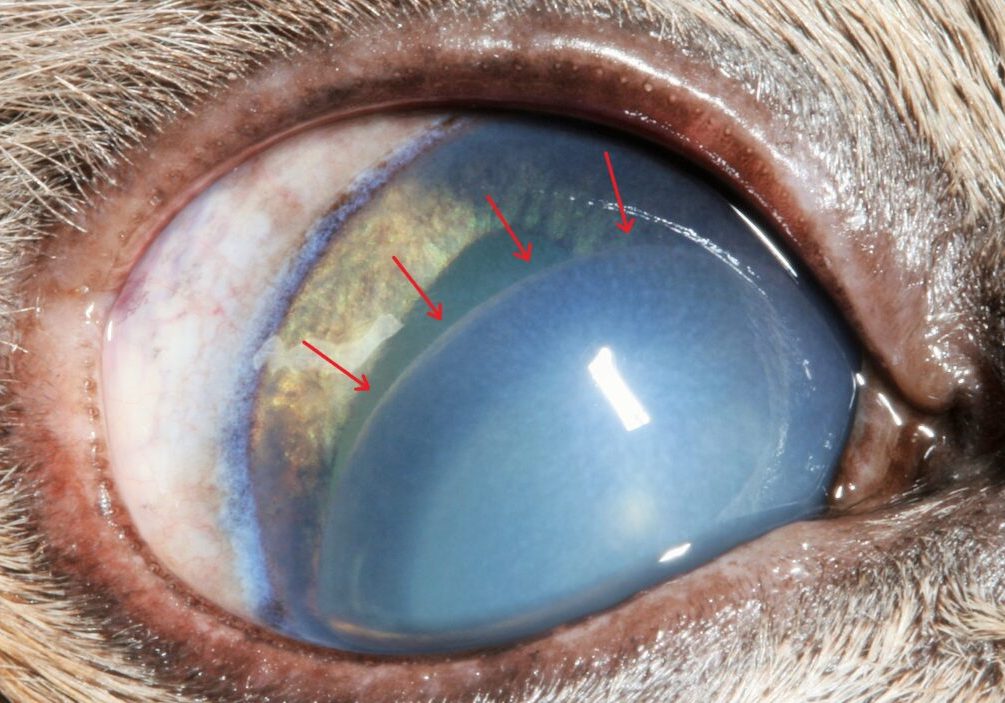
Fig 1: A dislocated lens in the front chamber of a cat eye. The arrows mark the edge of the lens.
What are the Signs of Lens Luxation?
If the lens becomes trapped in the front of the eye, it can cause sudden onset of eye pain, clouding of the cornea (seen as blueing of the front of the eye) and redness of the “white” of the eye. The patient is often depressed and reluctant to exercise.
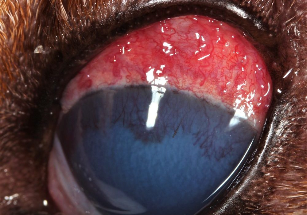
Fig 2: Lens luxation causing conjunctival redness and clouding of the cornea in a dog.
How is Lens Luxation Diagnosed?
Once the lens completely dislocates it is relatively straightforward to see that it is out of its normal position. A full ophthalmic examination by our Ophthalmology Specialist team using specialised equipment will confirm the diagnosis, and the most appropriate treatment will then be discussed.


What Treatments are Available for Lens Luxation?
Lens luxation is a serious, blinding and painful condition. In most cases, surgical removal of the lens is likely to give the best chance of preserving vision.
The surgical removal of a luxated lens is an operation that should only be carried out by an Ophthalmic Specialist due to the special training and skills required to perform the procedure. The surgery requires the use of a muscle-relaxant anaesthetic, to ensure the eye is held absolutely still and in the right position during the operation, our Specialist Anaesthetists ensure that the anaesthetic and the patients breathing is closely monitored.
Pic 3: A visual (seeing) eye following lens removal, showing a faint scar at the top of the eye.
What can I Expect if my Pet is Treated for Lens Luxation?
Most pets will cope very well without a lens but will take a little time to adapt to the new vision. Most dogs without lenses are able to navigate well avoiding obstacles, and many can still chase a ball.
The chance of a successful outcome depends in part on how long the underlying problem existed prior to presentation of symptoms. For patients who have been treated in the early stages the outcome of surgery can be very satisfactory, however in longstanding cases the success chances are significantly reduced.
Unfortunately, post-surgery blinding complications do occur depending upon the condition of the eye and duration of the problem at the time of surgery. These complications include persistence of glaucoma, bleeding into the eye and retinal detachment.
To help reduce the risk complications, six monthly check ups are recommend. In some cases, it may be necessary to continue with long-term medication, to keep the pressure in the eye under control.
In advanced cases of lens luxation, when the eye is painful and has lost vision permanently due to lens luxation, it may be necessary to consider removal of the eye under these circumstances.
Eye removal (enucleation) is generally only recommended if the eye is painful and blind and the pain cannot be readily controlled with medication. The thought of an eye being removed may be alarming at first, however it will generally make the patient feel much more comfortable and much happier very quickly so they can continue to enjoy life!
To save this page as a PDF, click the button and make sure “Save as PDF” is selected.
Ophthalmology
Find out more
To assist owners in understanding more about Ophthalmology we have put together a range of information sheets to talk you through the some of the more common conditions seen and treated by our Specialists.