Total Ear Canal Ablation Surgery
Why Should I Bring my Pet to Willows for Total Ear Canal Ablation Surgery?
Willows is one of Europe’s leading small animal referral centres. Our state-of-the-art hospital is led by internationally renowned Specialists, committed to providing the highest standards of veterinary care. Total Ear Canal Ablation Surgery is a complex procedure, our Soft Tissue Specialists have extensive experience in the successful completion of this intricate procedure.
Our team of Surgeons are supported by our multi-disciplinary team of Specialists across a number of disciplines including; Anaesthesia, Diagnostic Imaging and Emergency and Critical Care. Willows also has a large dedicated team of Vets, Nurses and clinical support staff available 24 hours a day, every day of the year to provide the best possible care for your pet.
What is a Total Ear Canal Ablation Surgery (TECA)?
Total Ear Canal Ablation is a procedure performed to manage severe canal or middle ear disease in dogs. The full name of the procedure is Total Ear Canal Ablation with Lateral Bulla Osteotomy (TECA + LBO) however is usually shortened to TECA for convenience.
A TECA involves the removal of the diseased and infected ear canal whilst leaving the inner ear (the hearing organ) itself (the inner ear) in place. The middle ear chamber (tympanic bulla) is carefully inspected and any abnormal tissue or material is removed.
Some useful definitions:
- Ear pinna: The visible part of a dog’s ear, the appearance varies between different breeds and can stick up or be floppy. Most dogs are able to swivel and move their pinna in order to pick up sounds around them.
- Ear canal: An air filled ‘trumpet’ shaped tube that allows sound waves to be channelled from the ear pinna to the ear drum.
- Ear drum: A very thin sheet of tissue that catches the sound waves and transmits them via three very small bones to the inner ear.
- Tympanic bulla: Known as the ‘middle ear’ it hosts the ear drum and acts as an amplifying chamber.
- Hearing organ: Receives the sound as vibrations and converts it into electrical signals that are then sent to the brain for interpretation. Situated within the skull, it is known as the ‘inner ear’.
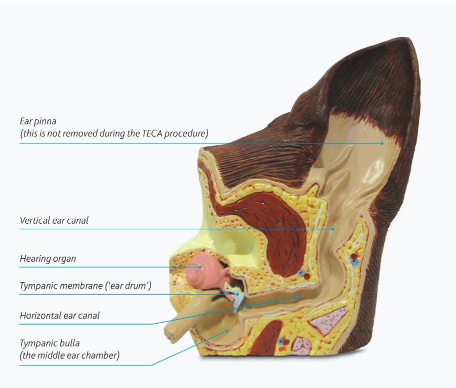
Fig1: Normal anatomy of the outer, middle and inner ear.
What are the Most Common Causes of Ear Disease in Dogs?
Canine ear disease usually occurs due to inflammation of the skin that lines the ear canal (otitis externa) which can lead to secondary infection of the middle ear chamber (otitis media).
In the majority of cases, inflammation of the skin within the ears is part of a more generalised skin condition, dogs suffering from ear problems often lick or chew at their feet or experience irritation elsewhere. More severely affected dogs can suffer from clear evidence of skin allergy over other parts of their body, in addition to the ear disease.
As infection and inflammation of the ear canal progresses it can become irreversibly narrowed, and the middle ear chamber can also become filled with infected material (Fig2).
Most cases of severe ear disease are assessed initially by our Specialist in Dermatology who will perform a full assessment of the ear and advise on potential treatment options.
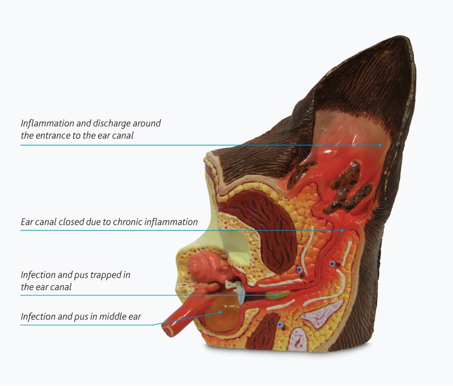
Fig 2: A model of an ear suffering from ear disease with middle ear involvement
What Treatments are Available for Ear Disease?
It is most appropriate to ensure that all reasonable steps have been taken to manage the condition medically before seeking a surgical solution for ear disease.
A Primary Care Veterinary Surgeon can often help you with this, alternatively our Specialist Dermatologist at Willows may offer additional conservative management options.
TECA surgery is reserved for those cases that cannot be managed satisfactorily by medical means.
Fig 3: A model of diseased ear and indicates the structures that are removed during TECA surgery
It is most appropriate to ensure that all reasonable steps have been taken to manage the condition medically before seeking a surgical solution for ear disease.
A Primary Care Veterinary Surgeon can often help you with this, alternatively our Specialist Dermatologist at Willows may offer additional conservative management options.
TECA surgery is reserved for those cases that cannot be managed satisfactorily by medical means.
What can I Expect if my Dog undergoes Total Ear Canal Ablation?
Whilst TECA surgery does not involve the removal of the inner ear/hearing organ, the removal of the ear canal itself will result reduced hearing sensitivity, similar to the effect of ear plugs or being under water.
When performed by a Specialist Surgeon, more than 90% of owners report a significant improvement in their dog’s quality of life following the procedure.
Whilst the vast majority of procedures result in success, there are some potential complications that can occur. Our Specialist Surgeons will take every step possible to minimise the risk of complications and maximise the chances of a good outcome.
Possible complications include:
- Reduced hearing sensitivity; similar to the effect of ear plugs or being under water
- Facial nerve paralysis; can occur 10-20% of cases and is usually temporary. The nerve runs very close to the ear canal and can be bruised during the procedure. Paralysis results in some mild drooping of the upper lip and eyelid on the affected side and can result in a reduced blink reflex. Eye lubrication may be required for a time until the third eyelid adapts. Most cases resolve over 4 to 6 weeks.
- Bleeding; significant bleeding can occur during the TECA operation due to the location of blood vessels. All possible steps will be taken to avoid bleeding and minimise the risk of life threatening levels of haemorrhage.
- Wound healing complications; patients will need to wear an Elizabethan collar following the procedure until the stitches are removed.
- Vestibular syndrome (balance problems); the organ responsible for balance is positioned next to the organ responsible for hearing. In some rare cases damage to this region is unavoidable due to the extent of the disease process. Dogs with the complication of balance dysfunction (similar to vertigo) often require significantly more nursing in the immediate postoperative period and some may never fully recover.
- Infection/abscess formation; when performed by Specialist Surgeons the occurrence infection or abscesses are is very low. Operating in such infected tissue can result in the risk of abscessation which can prove extremely difficult to resolve. Further surgery may be required and not all cases are successfully resolved.
- Horner’s syndrome; a combination of clinical signs that can result from damage to a complex of nerves that cross the middle ear. Damage to these nerves causes protrusion of the third eyelid, drooping of the upper eyelid and constriction of the pupil of the eye on the operated side. These signs are infrequent in the dog, and are usually temporary. Those cases that do not fully recover do not experience a significant reduction in quality of life or ability to see.
To save this page as a PDF, click the button and make sure “Save as PDF” is selected.
Soft Tissue Surgery
Find out more
To assist owners in understanding more about Soft Tissue Surgery we have put together a range of information sheets to talk you through the some of the more common soft tissue conditions seen and treated by our Specialists.
Condition:
Treatment: